Your Gastric antral vascular ectasia histology images are ready. Gastric antral vascular ectasia histology are a topic that is being searched for and liked by netizens now. You can Get the Gastric antral vascular ectasia histology files here. Get all royalty-free vectors.
If you’re looking for gastric antral vascular ectasia histology images information related to the gastric antral vascular ectasia histology interest, you have come to the ideal site. Our site frequently provides you with suggestions for viewing the maximum quality video and image content, please kindly hunt and locate more informative video content and images that match your interests.
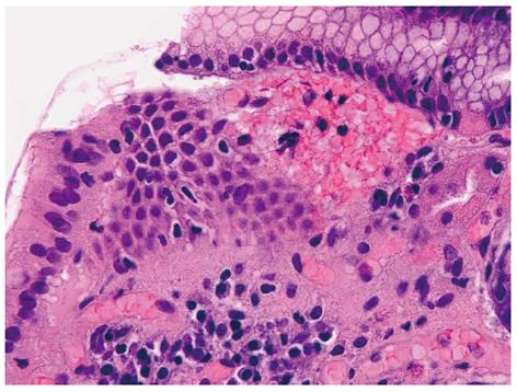
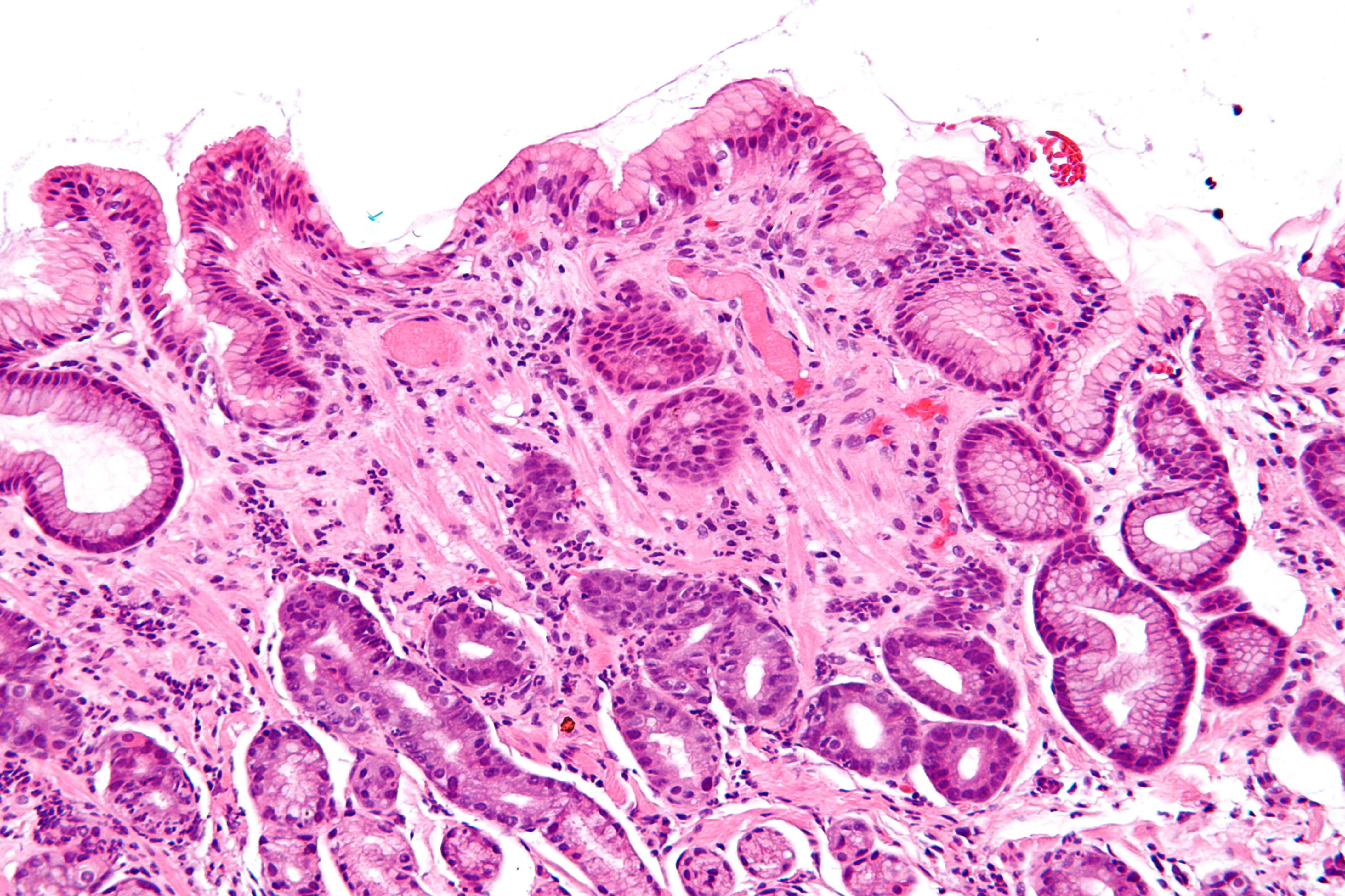
Gastric Antral Vascular Ectasia Histology. Fibrin thrombi in capillaries. The etiology of gastric antral vascular ectasia GAVE and hyperplastic polyps HP is not fully understood but there is no known overlap. A review of the pathogenesis and a comparative image analysis morphometric study of GAVE syndrome and gastric hyperplastic polyps. Associated with connective tissue diseases particularly systemic sclerosis but etiology unclear Arch Pathol Lab Med 2002126375 70 occur in elderly women.
 Hopkins Gi Pathology On Twitter Gastric Antral Vascular Ectasia Watermelon Stomach Dilated Vessels With Fibrin Thrombi Can Bleed Pathologists Http T Co Xipuazytyc Twitter From twitter.com
The red spots can merge causing stripes into the pylorus which has led to the term watermelon stomach. At endoscopy parallel longitudinal folds of gastric mucosa are. Also called watermelon stomach. Gastric antral vascular ectasia GAVE is the underlying cause for 4 of nonvariceal upper GI bleeding. A review of the pathogenesis and a comparative image analysis morphometric study of GAVE syndrome and gastric hyperplastic polyps. Gastroenterologists often biopsy the organ.
The etiology of gastric antral vascular ectasia GAVE and hyperplastic polyps HP is not fully understood but there is no known overlap.
It typically presents in middle aged females. It is often inflamed and may be a site that cancer arises from. A review of the pathogenesis and a comparative image analysis morphometric study of GAVE syndrome and gastric hyperplastic polyps. Mucosal vascular capillary ectasia. We included all cirrhotic. Gastric antral vascular ectasia GAVE is an uncommon but often severe cause of upper gastrointestinal GI bleeding responsible of about 4 of non-variceal upper GI haemorrhage.

It connects the esophagus to the duodenum. Gastric antral vascular ectasia GAVE is an uncommon but often severe cause of upper gastrointestinal GI bleeding responsible of about 4 of non-variceal up-per GI haemorrhage. Gastric antral vascular ectasia GAVE is an uncommon cause of occult gastrointestinal GI bleeding. The histology is characterized by marked dilation of capillaries and venules in the gastric mucosa submucosa with areas of intimal thickening spindle cell proliferation fibrohyalinosis and thrombi 4 10. 1 in 1953 refers to dilated blood vessels located in the antrum and radiated to the pylorus.
 Source: sciencedirect.com
Mucosal vascular capillary ectasia. Nodular GAVE and gastric hyperplastic polyps have similar appearance on upper GI endoscopy EGD as well as histology which could delay specific targeted therapy. 1 in 1953 refers to dilated blood vessels located in the antrum and radiated to the pylorus. It has been reported to account for 4 of all upper non-variceal gastrointestinal bleeding and up to 62 of patients afflicted with the disease become transfusion dependent 1 2. Gastric antral vascular ectasia GAVE is an uncommon cause of occult gastrointestinal GI bleeding.
 Source: wjgnet.com
Gastric antral vascular ectasia GAVE is an uncommon but often severe cause of upper gastrointestinal GI bleeding responsible of about 4 of non-variceal up-per GI haemorrhage. The diagnosis is mainly based on endoscopic pattern and for uncertain cases on histology. 1 in 1953 refers to dilated blood vessels located in the antrum and radiated to the pylorus. 2 in 1984 as its typical appearance is similar to the stripes seen on watermelons. Stomach is an important organ for pathologists.
 Source: sciencedirect.com
Gastric antral vascular ectasia GAVE also known as watermelon stomach WS is an uncommon cause of gastrointestinal GI blood loss. Gastric antral vascular ectasia GAVE also known as watermelon stomach WS is an uncommon cause of gastrointestinal GI blood loss. We performed this retrospective study at Centre Hospitalier de lUniversité de Montréal CHUM. Gastric antral vascular ectasia GAVE is a rare disorder characterized by its distinctive endoscopic appearance consisting of red tortuous ectatic blood vessels arranged in a longitudinal array localized in the gastric antrum converging on the pylorus. Nodular GAVE and gastric hyperplastic polyps have similar appearance on upper GI endoscopy EGD as well as histology which could delay specific targeted therapy.
 Source: researchgate.net
The diagnosis is mainly based on endoscopic pattern and for uncertain cases on histology. Gretz JE Achem SR. It was also named watermelon stomach by Jabbari et al. The term watermelon stomach was coined by Jabbari in 1984 to describe the endoscopic appearances2 In con-trast to the telangiectasia ofportal hypertension andOsler-Weber-Rendu there are nolesions in. It connects the esophagus to the duodenum.
 Source: commons.wikimedia.org
Gretz JE Achem SR. Gastric antral vascular ectasia or GAVE syndrome was first described by Rider et al in 195362 but was accurately defined by Jabbariet al in 198463 GAVE is characterised by red patches or spots in either a diffuse or linear array in the antrum. Gastric antral vascular ectasia GAVE is an uncommon cause of occult gastrointestinal GI bleeding. We included all cirrhotic. The diagnosis is mainly based on endoscopic pattern and for uncertain cases on histology.
 Source: youtube.com
Management of portal hypertensive gastropathy is centred on reduction in portal pres-sures whereas treatment of gastric antral vascular ectasia is predominantly endoscopic. Gastric antral vascular ectasia. Gastric antral vascular ectasia GAVE is an uncommon but often severe cause of upper gastrointestinal GI bleeding responsible of about 4 of non-variceal upper GI haemorrhage. Gastric antral vascular ectasia GAVE is the underlying cause for 4 of nonvariceal upper GI bleeding. Gastric antral vascular ectasia GAVE is an uncommon but often severe cause of upper gastrointestinal GI bleeding responsible of about 4 of non-variceal up-per GI haemorrhage.
 Source: semanticscholar.org
Gastric antral vascular ectasia watermelon stomach was first described in 1953 by Rider et all and about 50 further cases have been subsequently reported. We performed this retrospective study at Centre Hospitalier de lUniversité de Montréal CHUM. Gastric antral vascular ectasia GAVE is a disease process in the stomach typically presenting with chronic blood loss and iron-deficiency anemia. Am J Clin Pathol. Rare acquired vascular disease that may cause blood loss and iron deficiency anemia due to chronic antral hemorrhage.
 Source: twitter.com
Definition general. Nodular GAVE and gastric hyperplastic polyps have similar appearance on upper GI endoscopy EGD as well as histology which could delay specific targeted therapy. Gastric antral vascular ectasia GAVE is a disease process in the stomach typically presenting with chronic blood loss and iron-deficiency anemia. Introduction Gastric antral vascular ectasia GAVE is the underlying cause for 4 of nonvariceal upper GI bleeding. 2 in 1984 as its typical appearance is similar to the stripes seen on watermelons.
 Source: giejournal.org
Mucosal vascular capillary ectasia. Gastric antral vascular ectasia GAVE is an uncommon but often severe cause of upper gastrointestinal GI bleeding responsible of about 4 of non-variceal up-per GI haemorrhage. Submucosa may have tortuous vessels. It connects the esophagus to the duodenum. The histology is characterized by marked dilation of capillaries and venules in the gastric mucosa submucosa with areas of intimal thickening spindle cell proliferation fibrohyalinosis and thrombi 4 10.
 Source: twitter.com
Gastric antral vascular ectasia GAVE is an uncommon cause of occult gastrointestinal GI bleeding. 2 in 1984 as its typical appearance is similar to the stripes seen on watermelons. Nodular GAVE and gastric hyperplastic polyps have similar appearance on upper GI endoscopy EGD as well as histology which could delay specific targeted therapy. Introduction Gastric antral vascular ectasia GAVE is the underlying cause for 4 of nonvariceal upper GI bleeding. Watermelon stomach from the endoscopic appearance Diagnostic Criteria.
 Source: commons.wikimedia.org
Gretz JE Achem SR. Gastric antral vascular ectasia GAVE is an uncommon but often severe cause of upper gastrointestinal GI bleeding responsible of about 4 of non-variceal up-per GI haemorrhage. We report a case of gastroduodenal HP arising in a patient treated for GAVE. A review of the pathogenesis and a comparative image analysis morphometric study of GAVE syndrome and gastric hyperplastic polyps. Also called watermelon stomach.
 Source: semanticscholar.org
It is often inflamed and may be a site that cancer arises from. Introduction Gastric antral vascular ectasia GAVE is the underlying cause for 4 of nonvariceal upper GI bleeding. Associated with connective tissue diseases particularly systemic sclerosis but etiology unclear Arch Pathol Lab Med 2002126375 70 occur in elderly women. The diagnosis is mainly based on endoscopic pattern and for uncertain cases on histology. Gastric antral vascular ectasia GAVE also known as watermelon stomach WS is an uncommon cause of gastrointestinal GI blood loss.
 Source: commons.wikimedia.org
Gastric antral vascular ectasia GAVE or watermelon stomach has been recognised as a distinct pathological lesion since 19843 GAVE exclusively affects the antrum of the stomach. The red spots can merge causing stripes into the pylorus which has led to the term watermelon stomach. An introduction to gastrointestinal pathology is in the gastrointestinal pathology article. Watermelon stomach from the endoscopic appearance Diagnostic Criteria. Am J Clin Pathol.
 Source: europepmc.org
Management of portal hypertensive gastropathy is centred on reduction in portal pres-sures whereas treatment of gastric antral vascular ectasia is predominantly endoscopic. At endoscopy parallel longitudinal folds of gastric mucosa are. Vesoulis Z Naik N Maseelall P. Histopathologic changes are not specific for diagnosis of gastric antral vascular ectasia GAVE syndrome. Watermelon stomach from the endoscopic appearance Diagnostic Criteria.
 Source: commons.wikimedia.org
Gastric antral vascular ectasia GAVE is a disease process in the stomach typically presenting with chronic blood loss and iron-deficiency anemia. Gastroenterologists often biopsy the organ. Gastric antral vascular ectasia GAVE is an uncommon but often severe cause of upper gastrointestinal GI bleeding responsible of about 4 of non-variceal up-per GI haemorrhage. A review of the pathogenesis and a comparative image analysis morphometric study of GAVE syndrome and gastric hyperplastic polyps. Also called watermelon stomach.
 Source: pathologyoutlines.com
At endoscopy parallel longitudinal folds of gastric mucosa are. 1 in 1953 refers to dilated blood vessels located in the antrum and radiated to the pylorus. Gastric antral vascular ectasia or GAVE syndrome was first described by Rider et al in 195362 but was accurately defined by Jabbariet al in 198463 GAVE is characterised by red patches or spots in either a diffuse or linear array in the antrum. It was also named watermelon stomach by Jabbari et al. Distinctive pattern of gastric antral vascular changes with an associate distinctive endoscopic appearance.
 Source: librepathology.org
Gastric antral vascular ectasia GAVE is the underlying cause for 4 of nonvariceal upper GI bleeding. Nodular GAVE and gastric hyperplastic polyps have similar appearance on upper GI endoscopy EGD as well as histology which could delay specific targeted therapy. Gastric antral vascular ectasia GAVE also known as watermelon stomach WS is an uncommon cause of gastrointestinal GI blood loss. Am J Clin Pathol. Gastric antral vascular ectasia GAVE is a disease process in the stomach typically presenting with chronic blood loss and iron-deficiency anemia.
This site is an open community for users to do submittion their favorite wallpapers on the internet, all images or pictures in this website are for personal wallpaper use only, it is stricly prohibited to use this wallpaper for commercial purposes, if you are the author and find this image is shared without your permission, please kindly raise a DMCA report to Us.
If you find this site adventageous, please support us by sharing this posts to your preference social media accounts like Facebook, Instagram and so on or you can also bookmark this blog page with the title gastric antral vascular ectasia histology by using Ctrl + D for devices a laptop with a Windows operating system or Command + D for laptops with an Apple operating system. If you use a smartphone, you can also use the drawer menu of the browser you are using. Whether it’s a Windows, Mac, iOS or Android operating system, you will still be able to bookmark this website.


